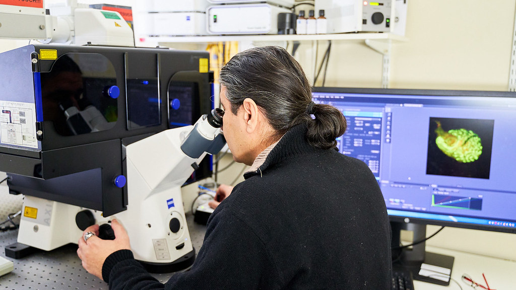
Our instruments form a part of a wider imaging network within Bath. Learn more about the Light Microscopy Research Network at Bath.
The Bio-Imaging and Cell Analysis Unit encompasses several different approaches to imaging biological and non-biological samples, the common denominator being the use of a light source, usually in the visible spectrum (photons), to produce a signal, that is then read by a suitable detector (our eyes or a specialised electronic device). This is in contrast to electron microscopy, where the "light" source is actually a beam of electrons.
Our bio-imaging instruments are employed in a variety of different projects, including studies of inflammatory diseases, cancer research, stem cell biology, diabetes, neuroscience, developmental biology, and drug discovery. Training and assistance are provided at all levels, including guidance on planning your imaging experiments and preparing your samples, and help with image and data analysis.
Light Microscopy
Light microscopy refers to all the techniques that employ a light source at the visible spectrum and a microscope, in order to view very small objects, usually < 1 mm. Today, there are two major types of light microscopy recognized: Brightfield and Fluorescent microscopy.
- Zeiss LSM880 confocal system with Airyscan & Multi-photon laser
- Zeiss Z.1 LightSheet
- Leica Dmi8 widefield system
The Confocal laser scanning microscopes enable researchers to create detailed 3D pictures of cell organelles and to examine ‘live’ cells through incubation systems that facilitate the study of cellular changes over time.
Read on to learn about Second Harmonic Generation microscopy.
Hypoxic Facility
With this facility we can control oxygen levels to study different aspects of cell function, from a single cell through to entire cell populations. This will provide cell models that mimic oxygen levels in living systems. We use these cell models to explore the effects of oxygen deprivation, which can lead to tissue damage and disease. Our equipment provides a complete and integrated solution for diverse hypoxic/anoxic studies:
- Baker Ruskinn Sci-tive dual hypoxic workstation
- Leica Dmi8 widefield microscope (housed inside the hypoxic workstation)
- BMG Labtech CLARIOstar plate reader (with regulated oxygen control)

