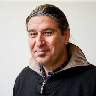
Michael Zachariadis Instrument Specialist - Bio Imaging
Michael manages the Bio Imaging instruments in the Imaging Facility, providing support, expert advice, and training in image and data acquisition and analysis.
Role
Michael’s primary role is the management of all Bio Imaging instruments in the Imaging Facility, including confocal and multiphoton microscopes, FACs analysers, plate readers, a high-content imaging system, and the hypoxic facility. This includes the day-to-day running and maintenance of the equipment, ensuring unhindered operation and high quality images and data.
In addition, Michael provides support and expert advice relating to Bio Imaging microscopy and microscopy/imaging in general, including:
- training of users
- optimising image and data acquisition
- helping with image and data processing and analysis
- developing new techniques and methods
Michael also helps with outreach activities; delivering seminars, training courses and specialist lectures on established and new techniques and capabilities to all researchers at Bath.
Career
Michael has more than 20 years' experience in the field of microscopy, and has worked with both light and electron microscopes. From 2016 to 2018 he managed the Confocal Imaging Unit at the Institute of Biosciences and Applications at N.C.S.R. "Demokritos" in Athens, Greece. His role included supporting the operation of a newly acquired Leica TCS SP8-MP multiphoton microscope, and providing expertise in the use of advanced imaging methodologies and techniques, as well as in image and data processing and analysis.
Previously, Michael worked in a Postdoctoral Research Associate capacity in the lab of Dr. Dedos, Faculty of Biology, University of Athens, Greece. He was part of a multidisciplinary team working to identify different modes of interaction between the inositol 1,4,5-trisphosphate receptor and adenylate cyclase 6, by combining molecular biology and microscopy techniques. His particular role was to set-up and implement novel imaging techniques, like FRET, PLA, Calcium Imaging and Live Cell Imaging.
In the past he has also managed an electron microscopy lab in the Department of Anatomy at the Medical School of Athens, Greece. Moreover, he has extensive teaching experience, both for undergraduates and postgraduates, has trained colleagues in microscopy, and supervised PhD students.
Education
Michael’s journey in Biology begun in 1992 when he started his studies at the University of Athens, Greece. He received his BSc in Biology in 1996 after completing his diploma thesis on the development of stomata in ferns, in the Department of Botany under the supervision of Prof Galatis. This work opened the door to the world of microscopy for Michael, who, in contrast to his peers, has always been fascinated by microscopes and of life beyond the limits of the human eye.
Thereafter, he pursued a PhD in Biological Sciences at the University of Athens, which he received with honours in 2004. His research involved the organisation of microtubules in dividing plant cells, and their relationship with the endoplasmic reticulum. His main tools were immunofluorescence and electron microscopy. During this time he also made his first steps in confocal microscopy at the Universität Hamburg, in collaboration with Prof Quader.

