Scientists from the University of Bath have helped to figure out why shoals of fish flash silver as they twist through the water by studying how the shiny silver cells are created in zebrafish.
In mammals, including humans, there is only one pigment cell-type; the melanocyte. These cells are usually black or brown, are responsible for colouring both skin and hair, and underlie the skin cancer melanoma.
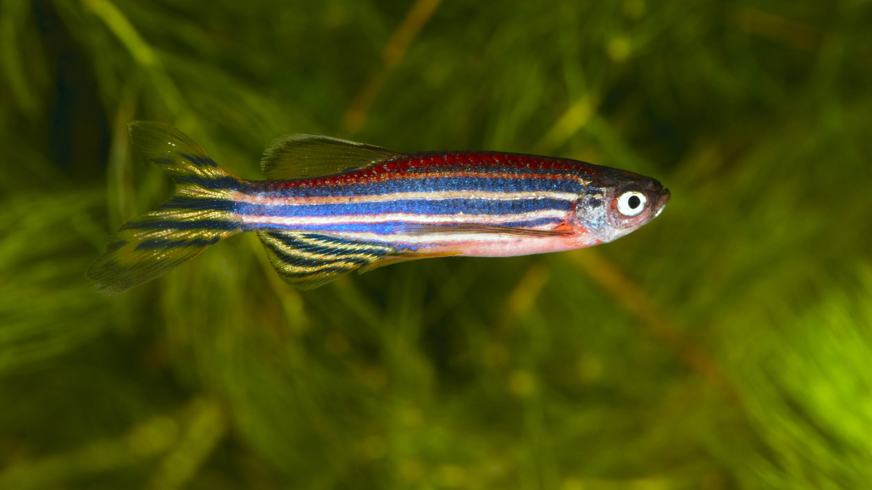
However fish form multiple pigment cell-types, not just the melanocytes. In zebrafish, in addition to black melanocytes, the body is coloured by yellow xanthophores, and by shiny, silver cells called iridophores.
The question of how different cells arise is a major question in stem cell and developmental biology, and this work from the laboratory of Prof. Robert Kelsh in the Department of Biology & Biochemistry sheds light on this question.
Prof. Kelsh’s group study the zebrafish, a small tropical fish, principally because they are easy to work with, are accessible for genetic manipulation, and have beautifully transparent embryos. This allows scientists to study cells of interest easily.
The different types of pigment cells derive from a type of stem cell called a neural crest cell, that also makes diverse types of neurons and skeletal cells, amongst others.
In this latest paper the lead author, Dr Kleio Petratou, and her colleagues used an unusual combination of genetic techniques and mathematical modelling to identify how a series of key genes interact and drive a neural crest cell to become a silver iridophore. The key genes were identified by genetic manipulation, demonstrating that when their function is lost, iridophores cannot be formed. But that didn’t explain how these genes worked together, a process made more difficult by the fact that neural crest cells migrate through the body extensively during the process of their development.
By using painstaking assessment of genetically-altered zebrafish and a rigorous focus on interpretation of the state of cells as they migrate, they were able to dissect the functional relationships between these genes, identifying what is known as a gene regulatory network (a kind of genetic wiring diagram) underpinning how a neural crest cell decides to become an iridophore.
Prof. Kelsh’s team used one other technique, mathematical modelling, to refine that network. Working with colleagues from the University of Surrey, they developed a series of simple mathematical models depicting alternative ways in which the network might be organised, and used computer simulations of their behaviour to identify a strongly favoured variant.
Dr Petratou said: “The combination of the sorts of detailed developmental genetics that the zebrafish allows, with rigorous mathematical modelling, really helped us discover the genes at the core of iridophore formation and to identify the structure of their interaction, in a way that simple genetics alone would not have allowed.”
Professor Kelsh added: “We are now in an excellent position to integrate this data with similar interdisciplinary studies on melanocytes, in order to understand how stem cell fate choice really works.” This approach has widespread application throughout stem cell biology, as well as showing how fish can shine. The study is published in PLOS Genetics.
The study was funded by the Biotechnology and Biological Sciences Research Council (BBSRC) and by University of Bath studentship to Dr Petratou.