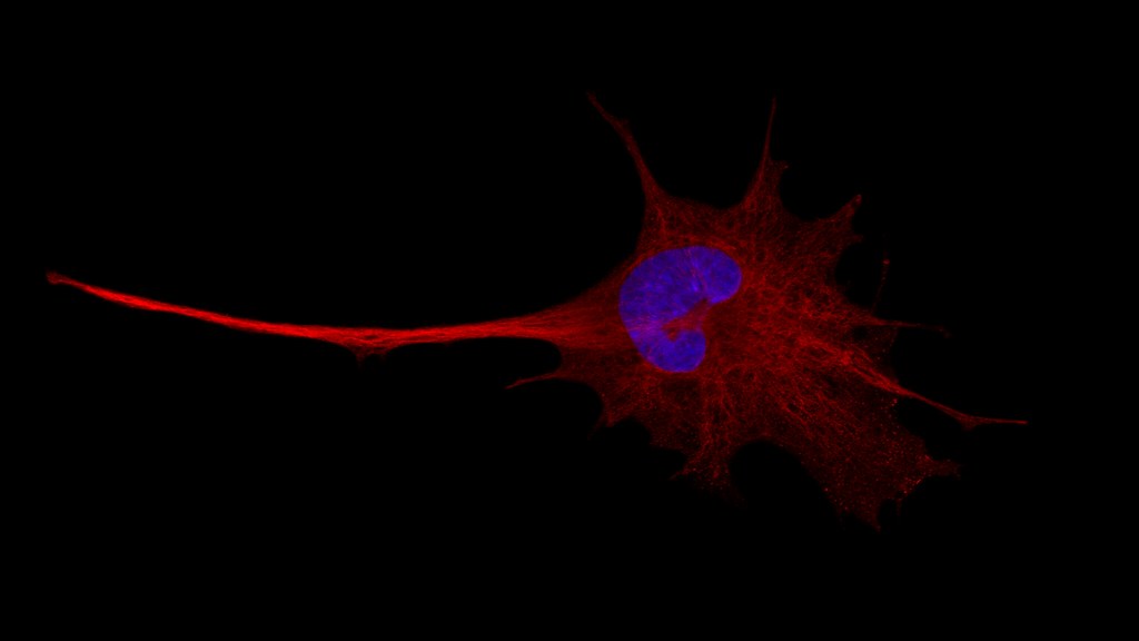
The Zeiss LSM-880 is a fully motorized, multi-modal inverted microscope system, based on a Carl Zeiss Axio Observer.Z1. The system offers single or two-photon excitation, spectral emission detection, multi-photon imaging, super resolution with an Airyscan detector, and full environmental control for Live Cell Imaging. The microscope is equipped with 7 excitation laser lines (405nm, 458nm, 488nm, 514nm, 561nm, 594nm, 633nm), as well as with an InSight DS+ Ti:Sapphire femtosecond pulsed laser (tunable between 690-1300nm), being able to excite the vast majority of the commercially available fluorophores. Depending on the application, emission is detected internally by a 34-channel detector array or the Super Resolution Airyscan detector, and externally by 2 PMTs at the transmitted path or a BIG.2 detector at the reflected path. A variety of air, oil and water objectives offer flexibility in imaging diverse samples. The offered imaging applications cover the biological, physical and material sciences including, but not limited to, multi-colour confocal imaging, multi-colour 3D imaging, 4D imaging (x, y, z, and time), live cell imaging, multi-photon (two-photon) imaging, Second/Third Harmonic Generation, steady-state Förster/fluorescence resonance energy transfer (FRET), fluorescence recovery after photobleaching (FRAP), spectral unmixing and fingerprinting, co-localisation studies, calcium imaging or other ratio imaging.
Funding details: Application of bioimaging and fluorescence-based bioprobes to study molecular and cellular mechanisms in integrated physiological settings. Wellcome Trust (2005) REF: 077454. PI: Prof. Stephen Ward.
